RISING STARS
A look at the exciting research going on in the labs
of Eric Yttri, Huaiying Zhang and Leon Zhao
by Jocelyn Duffy
In recent years, the Department of Biological Sciences has recruited a number of young faculty who are rising stars in their fields, undertaking exiting research that will impact science well into the future. Three of these junior faculty, Assistant Professors Eric Yttri, Huaiying Zhang and Yongxin “Leon” Zhao recently received grants to support their work.
ERIC YTTRI
The Yttri lab focuses on how the brain translates thought into action. Specifically using behavioral, physiological and computational methods to sample and manipulate the neural activity of multiple areas in the brain to understand the mechanisms behind behavior and movement diseases.
This fall, Yttri received a seed grant from the Brain Research Foundation to incorporate value in his experiments on behavior.
“We are trying to expand from not just understanding how we do something but why we do something and what motivates that. This project will help us to get a more complete picture of everything that goes into the transformation from thought to action, the motivation behind the choice and performance of behavior,” said Yttri.
Yttri is also developing tools that will advance his and other neuroscientists’ work. Yttri has teamed up with Associate Professor of Mechanical Engineering’s Rahul Panat to develop a new manufacturing method based on 3D nanoparticle printing to fabricate neural probes. Their technology, initially funded by a grant from the DSF Charitable Foundation, is now supported by a $1.95 million grant from the National Institutes of Health’s (NIH) Brain Research through Advancing Innovative Neurotechnologies (BRAIN) Initiative.
Yttri and Panat’s new 3D nanoparticle printing technology will create custom arrays that have a higher density of electrodes, allowing researchers to achieve the higher level resolution needed for applications such as precision neuroprosthetics. The electrode arrays created using their method will also be more durable and less expensive.
“With fMRI we can see the whole brain, but the temporal and spatial resolution are not where we need them to be. Electrodes can give us millisecond, single neuron resolution, but even with the most recent advances you might only be able to get information from 300 or 400 neurons at a time,” said Yttri. “By 3D printing the electrodes with our high throughput method, we can put the recording sites as close together or as far away as we want. And the nature of the electrode’s structure means they can be implanted in the brain much easier and with less damage than the current state of the art techniques.”
The researchers’ long-term goal is to create precision medical devices, such as brain machine interfaces. For example, a patient who needs an electrode for a neuroprosthetic could be given a device that, using structural MRI, could be customized based on maps of the individual curves of the brain.
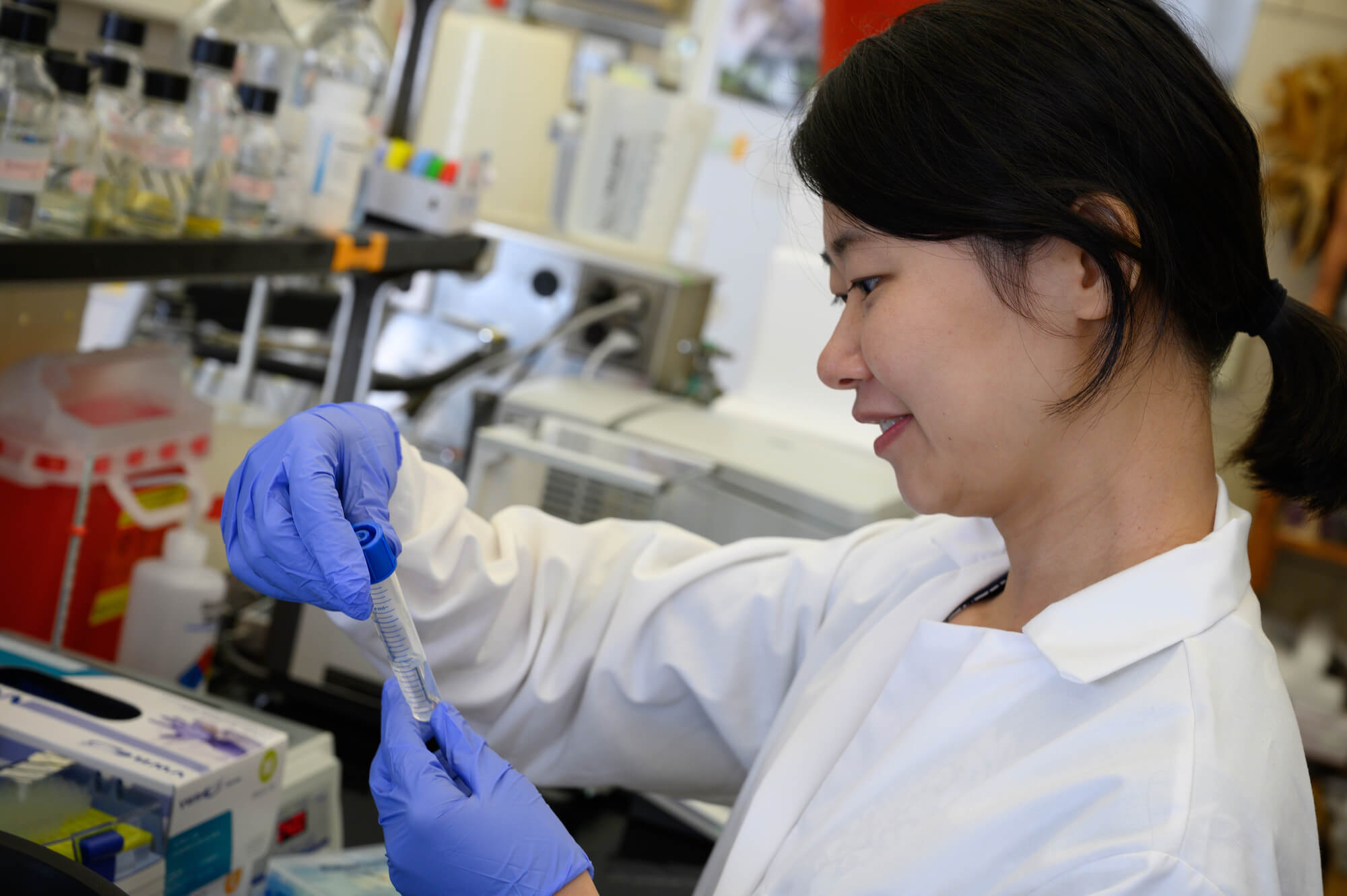
HUAIYING ZHANG
A biologist, Zhang’s work focuses on the physics and chemistry of liquid condensation in live cells, which could inform the development of new treatments for cancer. She received a New Investigator Award from the Charles E. Kaufman Foundation to support this work.
Human cells are organized into functional compartments called organelles. Most organelles, like the nucleus and mitochondria, are encased by lipid membranes. Others, like stress granules and nucleoli, do not have a membrane. In the last decade, researchers have discovered that these membrane-less organelles are condensed liquid droplets formed by liquid-liquid phase separation.
Zhang has developed optogenetic tools that control genetically engineered proteins using light to study how condensation occurs within the complex cellular environment and the role it plays in cellular function. Specifically, Zhang uses these tools to study membrane-less organelles called APBs that are associated with telomeres in some cancer cells. Telomeres, the protective endcaps of chromosomes, play an important role in cancer. In normal cells, telomeres shorten over the cells’ life span and when they reach a certain length, they trigger cell death. In cancer cells, telomeres maintain their length, allowing the cancer cells to live indefinitely.
“We found that APB assembly follows liquid condensation; so to dissect the roles of APBs in telomere elongation and eventually design cancer therapies targeting ABPs, we need to first understand the condensation process,” said Zhang.
Under the Kaufman grant, Zhang will attempt to determine the physics that underlies APB condensation and the chemistry behind the formation of APB and its material properties. Her interdisciplinary research stands to reveal insights into the foundational physics and chemistry of phase separation in live cells. It may also yield important information that will allow researchers to manipulate conditions within the cell to determine how APB condensation and properties can be altered to prevent telomere elongation, which could lead to new avenues of research in cancer therapy.
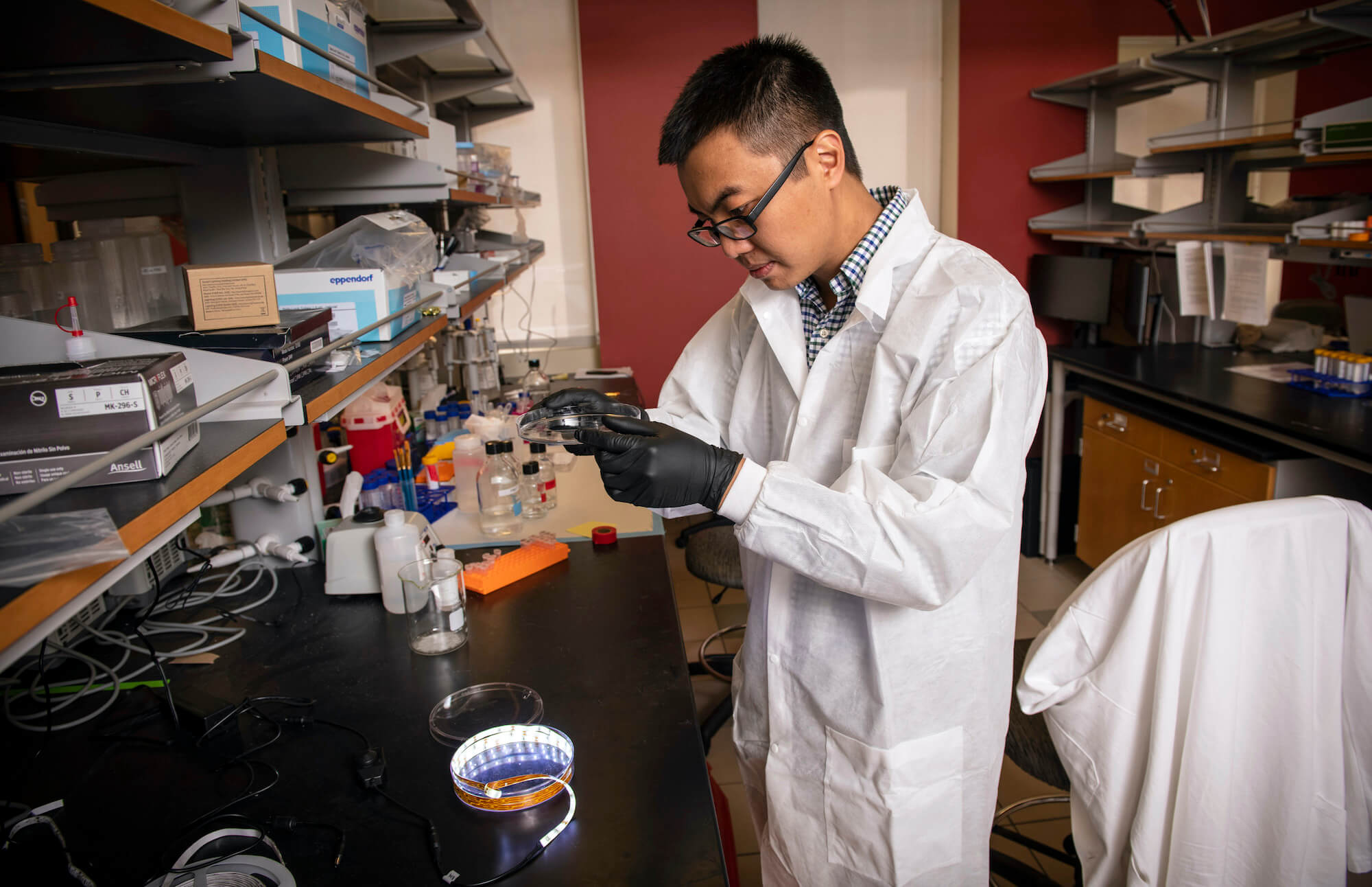
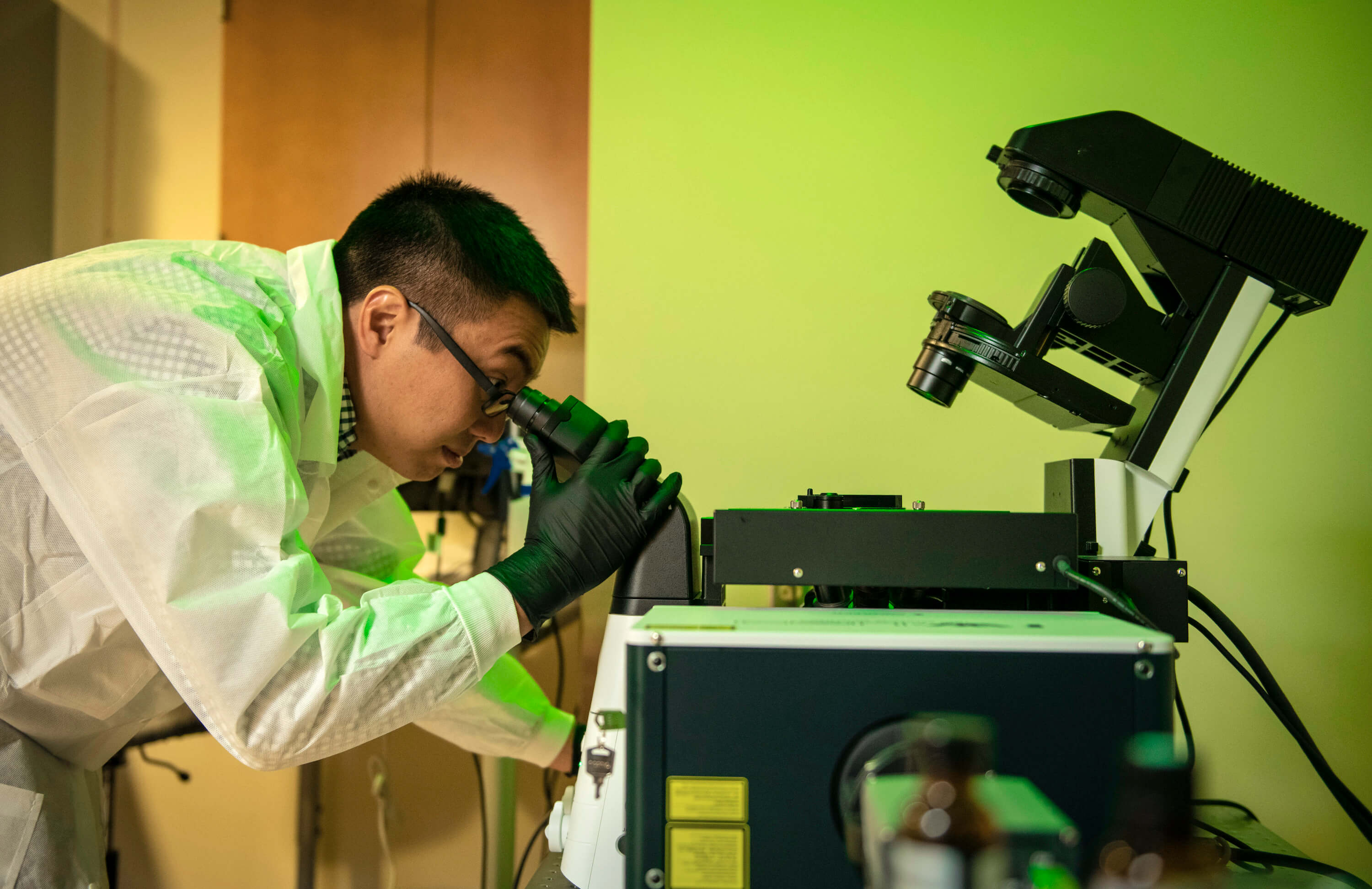
YONGXIN “LEON” ZHAO
Our bodies are made of molecular building blocks such as proteins, nucleic acids, lipids and carbohydrates that are assembled with nanoscale precision. Misconfiguration of these building blocks is a tell-tale sign of many diseases. Being able to characterize these misconfigurations at the nanoscale will lead to a better understanding of how disease progresses and will help with diagnostics and the development of new treatments.
Modern pathology traditionally uses regular light microscopy to analyze biopsies. This type of microscopy isn’t powerful enough to image biopsies at the nanoscale. To overcome this limitation, Zhao created a new technique called Expansion Pathology, which physically magnifies biopsies, allowing subtle pathological changes to be seen using existing microscopy equipment.
Zhao will use NIH funding to build on Expansion Pathology and create new chemistry and imaging strategies for more informative pathology at the nanoscale. Specifically, he will develop a portfolio of more powerful imaging platforms that allow for the direct observation of detailed biomolecule maps in biopsies. He plans to demonstrate the power of these platforms on a range of diseased tissues from precancerous breast lesions to infected tissues.
“We propose to physically expand biopsies to see more, departing from decades of practice in pathology that relies primarily on optical resolution,” said Zhao. “We hope that the new imaging techniques can find broad applications in pathology and medicine and elucidate the pathogenesis of many complex diseases, such as cancer, brain disorders and infectious diseases, fostering new discovery of biomarkers that enable accurate diagnoses and better therapeutics.”